Plastids:
Plastids are cytoplasmic organelles about 5 micron in diameter and 3 micron in thickness, found freely in cytoplasm of the most of plant cell. they are not found in animal cell. the term plastid was first used by Schimper (1885). They are usually disc shaped and arise from small granular bofies, the protoplastids. Based on the types of pigments, three types of plastids are recognized: leucoplast, chloroplast and chromoplast. One form of plastid can change into another form, for example, in tomatoes the ovary contains leucoplasts which change into chloroplasts in young fruits and finally into chromoplasts as the fruit ripens.
1)Leucoplasts: [Gr. leukos: white; plastikos: formed] These are colourless plastids found in non-green parts of the plant such as under-ground stem, root and meristemic cells. They may be rod-like, spherical or oval in shape. Each leucoplast is bound by double layered membrane which encloses granular matrix. They contain few lamellae and on exposure to sun light change into chloroplast by developing thylakoid structure. Leucoplasts are mainly concerned with storage of various kinds of reserve food materials and are named variously such as amyloplasts (involved in storage of starch), elaioplasts or lipoplast (involved in storage of oil), aleuroplasts (involved in storage of proteins).
2)Chloroplasts: [Gr. cholors: grass green] Chloroplasts were first observed by Antony Von Leewenhoek in 1679. These are green plastids found in green parts of the plan t exposed in sunlight. They are small, gren, discoid or ellipsoidal, 4 to 10 micron in diametr and 1 to 3 micron in thickness. They are abundantly found in leaf parenchyma. A leaf parenchyma may contain 20 to 40 chloroplasts in each cell. Each chloroplast is surrrounded by double layered membrane. The membrane is made up of lipids and proteins. It encloses granular matrix or stroma. The stroma contains double membrane lamellae, which run parellel to one another along the length of the chloroplast. The lamellae are also known as thylakoids. At places the lamellae form disc-shaped structures: the grana. In grana the lamellae are placed compactly one above the other like a stack of coins. The lamellae which are found in grana region called grana lamellae while the rest lamellae are designated as stroma lamellae or intergrana lamellae. The grana contain photosynthetic pigments, chlorophylls and carotenoids.
 t exposed in sunlight. They are small, gren, discoid or ellipsoidal, 4 to 10 micron in diametr and 1 to 3 micron in thickness. They are abundantly found in leaf parenchyma. A leaf parenchyma may contain 20 to 40 chloroplasts in each cell. Each chloroplast is surrrounded by double layered membrane. The membrane is made up of lipids and proteins. It encloses granular matrix or stroma. The stroma contains double membrane lamellae, which run parellel to one another along the length of the chloroplast. The lamellae are also known as thylakoids. At places the lamellae form disc-shaped structures: the grana. In grana the lamellae are placed compactly one above the other like a stack of coins. The lamellae which are found in grana region called grana lamellae while the rest lamellae are designated as stroma lamellae or intergrana lamellae. The grana contain photosynthetic pigments, chlorophylls and carotenoids.
On dry weight basis, the chloroplast shows following average chemical composition. Protein 40 to 50%, phospholipids 23-25%, chlorophyll 5-10%, carotenoids 1-2%, RNA 5% and DNA in small amount. Mg is present in about 2-3% of the total ash content. Fe and Cu are found in traces. Due to the presence of photosynthetic pigments the chloroplast forms the main centre of food synthesis. They trap solar energy and form food materials from CO2 and H2O. Chlorophyll provides green colour to the plant body.
3) Chromoplasts: [Gr: chroma - colour] These are coloured parts of plant such as flowers, fruits, etc. Their coloured pigments: carotenes (red), xanthophyll (yellow), etc are mostly present in vacuolar sap. They may arise from chloroplasts, as in petals and in pricarp of fruits or from leucoplasts in carrot roots. Chromoplast may also contain anthocyanin, a group of colouring matter dissolved in the vacuolar sap of petals which provides violet, purple, blue, brown and often red colour to the flowers. Chromoplasts are also bounded by double layered membrane which encloses matrix or stroma, but there is no lamellae and grana. Chromoplasts make flowers and fruits showy and attractive. The flowers attract insects and other animals, which help in pollination and dispersal of fruits and seeds.
Cilia and Flagella:
Cilia and flagella are thin projections from the surface of some cells. The y are identical structures. The name cilia is used for relatively short structures about 5 to 10 micron. They usually occur in large number and may cover the entire surface of the cell. The flagella are longer upto 150 micron, only one or two usually occuring on a cell. Cilia and flagella are usually not found in plant cells. However, the cells of some algae, and the male gametes of algae, bryophytes and pteridophytes possess cilia. In animal cells, they are most common in protozoans, sponges and other metazoa. A cilium or flagellum consists of a pair of central filaments surrounded by a ring of nine pairs or doublets peripheral filaments. They are bounded by an extension of cell membrane. Each cilium or flagellum arises from a basal body. The basal body remains embedded in the ectoplasm just below the surface of the cell. The basal body contains nucleic acids (DNA and RNA) and protein.
 y are identical structures. The name cilia is used for relatively short structures about 5 to 10 micron. They usually occur in large number and may cover the entire surface of the cell. The flagella are longer upto 150 micron, only one or two usually occuring on a cell. Cilia and flagella are usually not found in plant cells. However, the cells of some algae, and the male gametes of algae, bryophytes and pteridophytes possess cilia. In animal cells, they are most common in protozoans, sponges and other metazoa. A cilium or flagellum consists of a pair of central filaments surrounded by a ring of nine pairs or doublets peripheral filaments. They are bounded by an extension of cell membrane. Each cilium or flagellum arises from a basal body. The basal body remains embedded in the ectoplasm just below the surface of the cell. The basal body contains nucleic acids (DNA and RNA) and protein.
Functions:
1) They are locomotary organs and provide locomotion to the cell or organism.
2) The cilia create food currents in lower aquatic animals.
3) The ciliary movements help in the elimination of the solid particles from respiratory tract.
The flagella usually deat independently by unsulations forming a wave pattern. While the cilia move in a coordinated manner or rowing pattern, so that the overall effect is like a field of grass blowing gently in the wind..
Nucleus:
Nucleus was first developed by Robert Brown (1831) in an Orchid cell. It is a dense protoplasmic body lies embedded in the cytoplasm. It directs and controls all the activities of a cell. Usually the nucleus is spherical or oval in shape. The higher organism cell always contains a single nucleus in each cell, while the cell of some algae and fungi contain numerous nuclei. A well organised nucleus with a distinct nuclear membrane is present in all living cells except the prokaryotic cell or bacteria and blue-green algae. Usual size of nucleuus varies between 5 to 25 microns. In young meristematic cell, the nucleus lies in the centre, while in mature plant cell it lies in parietal layer of the cytoplasm or in the centre of the vacuole suspended by means of cytoplsmic strands. A nucleus always arises by the division of the pre-existing nucleus.
 Structure: A typical nucleus is composed of four parts: Nuclear membrane, Nucleoplasm, Nucleolus and Chromatin network.
1) Nuclear membrane: It is a double layered porous covering of the nucleus. It seperates the nucleus from the surrounding cytoplasm. The membranes are lipoproteinaceous in nature. The outer membrane at places gives out tubular structure, the endoplasmic reticulum. Nuclear membrane acts as a selective membrane and help in the movement of specific substances between the cytoplasm and the nucleus.
2) Nucleoplasm: The nuclear membrane encloses a clear structureless granular semi-fluid substance, the nucleoplasm. It is termed variously as nuclear sap or nuclear matrix or nucleoplasm or karyolymph. It is ismilar to the cytoplasm and additionally contains several enzymes which catalyse the synthesis and hydrolytic breakdown of nucleic acids and proteins.
3) Nucleolus:It is a small dense rounded body that remains attached to the chromatin. Usually a nucleus contains one nucleolus, whereas in some cases two or more nucleoli are found in a nucleus. Nucleolus takes part in synthesis of RNA are utilised in the formation of ribosomes. Nucleolus also forms spindle fibres during cell division.
4) Chromatin Networks: Nucleoplasm contains a number of dark staining fine thread-like structures called chromatin threads. The chromatin threads are actually elongated chrmosomes. They are coiled upon themselves and cross one another and form a tangled mass of threads, like a network, which is often called chromatin reticulum or network. Chromatin threads are the sites of main genetic material which control all the activities of the cell, metabolism and heredity. During cell division, these threads are separate, tightly coiled and condense to form short and thick structures, the chrmosomes.
Functions:
1) Nucleus directs and controls all the vital activities of a cell.
2) It contains hereditary information essential for continuity of species. It transmits all the hereditary information from generation to generation.
3) All instructions for reproduction, development, metabolism and behaviour are in the chromosome of nucleus.
4) Nucleoplasm contains many essential enzymes necessary for biochemical activities of the cell.
5) The nucleolus forms spindle fibres during cell division.
6) The nucleolus synthesises ribosomes and r-RNA fir the cell.
Vacuoles:
The fluid filled spaces within the cytoplasm are known as vacuoles. In young plant cell, the protoplasm fills the entire space of the cell, but as the cell grows, the protoplasm fails to keep pace with the incresing volume of the cell and causes numerous smaal spaces, the vacuoles. Finally, when the cell attains its maximum size these vacuoles are fused to form a large central vacuole occupying the whole central part of the cell. Thus, the protoplasm is pressed against the cell wall in the form of a thin layer. In animal cell, the vacuoles are either small or absent. Each vacuole is surrounded by a single membrane known as tonoplast. It is similar to the plasma membrane in composition and appearance. The vacuoles are filled with a watery fluid called the cell sap which contains many soluble inorganic and organic substances. Due to presence of these substances the sap of vacuoles possess high osmotic pressure, which has a great physiological significance. Secondly, the vacuole is thought to act as a reservoir for products of cell membrane. In many cases the sap also contains pigments known as anthocyanins and anthoxanthins. These pigments provide colouration to the various plant parts such as flowers, roots and stems. In many unicellular organisms, vacuoles help in getting rid of extra-water. for example: protozoans.
Chromosomes:
[Gr. Chroma: colour; soma: body] The rod-like chromosomes were observed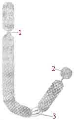 by Karl Nagli (1842) in the nuclei of plant cells. Starsburger (1875) discovered thread-like structures which appeared during cell division. The chromosomes are defined as the coloured body of nucleus. During metaphase and anaphase of cell division, the chromosomes become short and compact bodies having definite shape and size.
 by Karl Nagli (1842) in the nuclei of plant cells. Starsburger (1875) discovered thread-like structures which appeared during cell division. The chromosomes are defined as the coloured body of nucleus. During metaphase and anaphase of cell division, the chromosomes become short and compact bodies having definite shape and size.
Chromosomes's Number: The number of chromosomes for an organism is usually fixed, but it varies considerably among species. For example, a certain nematode worm species has only two chromosomes in each cell, while certain protozoa have over 300 chromosomes in each cell. A potato has 48, carrot has 18, corn has 20 and onion has 16 chromosomes in each cell.
Chromosomes regularly occur in pairs in all sexually reproducing organisms, one of which comes from the mother and the other from the father. The two members of each pair are called homologous chromosomes. Each chromosome of a pair has the same size and shape as its homologue. A cell with both the members of homologous pair is termed as diploid. The diploid number of chromosome is represented by 2n. The cell with only one chromosome of each homologous pair is called haploid. The haploid number of chromosome is represented by n. For example: a potato has 48 chromosomes, so there is 24 homologous pairs of chromosomes. The size of the chromosomes vary from 0.2 to 50 micron in length and 0.2 to 20 micron in diameter. the plant has generally larger sized chromosomes compared to that of the animals. the largest chromosomes are polytene and lampbrush chromosomes. The polytene chromosomes are formed by the repetition or duplication of the chromonema. They measure upto 2000 micron in length. It is mainly found in the tissues of the salivary glands, gut, trachea, fat body and Malpighian tubules of many insects of the order Diptera.
The lampbrush chromosomes are composed of main axis and the lateral loops. The chromonema of the chromatids forms fine loops at the lateral sides giving the appearance of lampbrush or test-tube brush. The length of the lampbrush chromosomes reach upto 5900 micron or three rimes longer than the polytene chromosomes. It occurs in yolk rich oocytes of many vertebrates such as fish, amphibians, reptiles and birds.
Structure: Structurally the chromosomes are rod-shaped, bounded by a proteinaceous pellicle which encloses a jelly-like substance called matrix. Each chromosome consisits of two distinct regions: Heterochromatin and Euchromatin.
During metaphase a chromosome appears to consists of two threas like structures called chromatids. These chromatids are attached together by a centromere. The chromatids consist of several thin filamentous chromosomal material, the chromonemata. These chromonemata are coiled together and enclosed in a separate common matrix to form a chromatid. At places, the group of chromonemata show maximum coiling to form dark band like structure called chromomere and the regions in between two chromomeres are known as interchromomeres.
Depending upon the position of centromere, the chrmosomes show different shape and are classified as: Telocentric, Acrocentric, Sub-metacentric, metacentric.


0 comments :
Post a Comment