The name earthworm is given to the annelid  worms, because they live and burrow in the ground. Annelids are triploblastic, bilaterally symmetrical and coelomate animals. The presence of true segmentation (metamerically segmented) in their body is a diagnostic feature of these animals. The locomotary organs are setae or chaetae present in each segment. Phylum Annelida, also sometimes known as Annulata, contains elongated, vermiform and true segmented animals.
Systematic Position:
Phylum: Annelida
Class: Oligochaeta
Order: Opisthopora
Genus: Pheretima
Species: posthuma
Habit and Habitat: Pheretima is commonly called earthworm. It is worldwide in distribution. They are found in the soil rich in decaying organic matters usually in lawns, gardens, pastures, near the banks of ponds, lakes and rivers. It generally inhabits in the upper layer of earth upto a depth of 12-18 inches. It is nocturnal in habit, i.e. it prefers to live in the burrow during the day and comes out at night. In damp cloudy weather after a heavy rain fall, the earthworms are seen in large numbers crawling on the ground. They make their burrow usually in moist soil, partly by boring with its pointed anterior end and partly by sucking and swallowing the soil. They feed on dead organic matter present in the soil. The soil is also ingested along with the food and is egested with undigested food in the form of worm castings.
External Morphology:
1) Shape and Size: The body is long, cylindrical narrow bilaterally symmetrical, nearly circular in cross section. The anterior end is tapering while the posterior end is more or less blunt. A mature earthworm measures about 150 mm in length and 3 to 5 mm in thickness. The thickest part of the body is little behind the anterior end. The dorsal surface is marked by the presence of a dark median line of dorsal blood vessel which runs along the length of the body just below the skin. Similarly the ventral surface is marked by the presence of genital openings and papillae in the anterior part of the body. The body is shiny dark-brown in colour. The dorsal surface is darker than the ventral surface. Brown colour of worm is due to the pigment called porhyrin present in body wall.
a) Segmentation: The body consists of about 100-120 segments. The segmentation is true, i.e. the external segmentation corresponds with internal segmentation of the body. Hence, the segmentation of Earthworm is known as metameric segmentation or metamerism. The whole body surface is divided by a distinct series of circular grooves. All the segments are alike except the first and the last.
b) Peristomium: They have no distinct head, eyes and tentacles. The first segment of the body is called peristomium or the buccal segment, which bears a terminal, crescentric mouth. It is prolonged anteriorly into a fleshy lobe, the prostomium, which overhangs the mouth.
c) Clitellum: A girdle-like thick band of glandular tissue is found towards the anterior end of the body, which is called the clitellum or cingulum. The clitellum is formed in mature worms and extends over 14th-16th segments. The glands of clitellum secrete mucus, albumen and egg case or cocoon for the eggs. Due to the presence of clitellum, the body of earthworm is divided into a pre-clitellar, clitellar and post-clitellar regions.
2) Apertures:
Mouth: It is a cresentric opening situated below the prostomium and surrounded by the first anterior segment of the body called the peristomium or the buccal segment.
Anus: It is a vertical slit-like opening present in the last segment of body. The last segment with anus is called the anal segment. It is the exit if the alimentary canal through which undigested wastes are discharged.
Genital Openings: Earthworm is a hermaphrodite animal, therefore the male and female genital apertures are found in the same individual. Male genital opening: A pair of crescentic openings are present on the ventral surface of the 18th segment, one on either side. Through these openings male reproductive bodies are discharged. Female genital opening: A single median aperture of the oviducts is present on the ventral surface of the 14th segment in a saucer-shaped depression. The female reproductive bodies are discharged through it.
Spermathecal Aperture: Four pairs of small ventrolateral spermathecal apertures are situated intersegmentally between the grooves of 5/6, 6/7, 7/8 and 8/9 segments. They store sperm during copulation.
Nephridiopores: A large number of very minute pores scattered irregularly all over the body surface, except the first two segments. These are the openings of the integumentary nephridia by which the metabolic wastes of the body are removed.
Dorsal Pores: Along the mid-dorsal line a series of minute openings are in the inter segmental groove. Through these dorsal ores are found behind the 12th segment except the last groove. Though these dorsal pores coelom communicates with the exterior. The coelcemic fluid oozes out through these pores and kills bacteria of the soil, it also keeps skin moist for respiration.
Genital Papillae: There are two pairs of circular and raised papillae present on the ventral surface of the 17th and 19th segments. These are present in the same line with the male genital openings, each of which bears a shallow cup-like depression at its tip. The genital papillae acts as suckers during copulation.
Setae: It is a chitinoid structure of a faint yellow colour and shaped like an elongate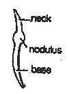 d "S". It has swollen middle part, the nodulus. except the first, the last and clitellum, each segment contains a ring of about 80 to 120 chitinous setae along its middle line. Such an arrangement is known as perichaetine arrangement. About one-third of setae length projects above the surface of skin, the remaining being embedded in a setal sac. The setae helps in locomotion by holding the earth since they are directed backwards. Their movement is controlled by special types of muscles.
3) Body Wall:
 worms, because they live and burrow in the ground. Annelids are triploblastic, bilaterally symmetrical and coelomate animals. The presence of true segmentation (metamerically segmented) in their body is a diagnostic feature of these animals. The locomotary organs are setae or chaetae present in each segment. Phylum Annelida, also sometimes known as Annulata, contains elongated, vermiform and true segmented animals.
Systematic Position:
Phylum: Annelida
Class: Oligochaeta
Order: Opisthopora
Genus: Pheretima
Species: posthuma
Habit and Habitat: Pheretima is commonly called earthworm. It is worldwide in distribution. They are found in the soil rich in decaying organic matters usually in lawns, gardens, pastures, near the banks of ponds, lakes and rivers. It generally inhabits in the upper layer of earth upto a depth of 12-18 inches. It is nocturnal in habit, i.e. it prefers to live in the burrow during the day and comes out at night. In damp cloudy weather after a heavy rain fall, the earthworms are seen in large numbers crawling on the ground. They make their burrow usually in moist soil, partly by boring with its pointed anterior end and partly by sucking and swallowing the soil. They feed on dead organic matter present in the soil. The soil is also ingested along with the food and is egested with undigested food in the form of worm castings.
External Morphology:
1) Shape and Size: The body is long, cylindrical narrow bilaterally symmetrical, nearly circular in cross section. The anterior end is tapering while the posterior end is more or less blunt. A mature earthworm measures about 150 mm in length and 3 to 5 mm in thickness. The thickest part of the body is little behind the anterior end. The dorsal surface is marked by the presence of a dark median line of dorsal blood vessel which runs along the length of the body just below the skin. Similarly the ventral surface is marked by the presence of genital openings and papillae in the anterior part of the body. The body is shiny dark-brown in colour. The dorsal surface is darker than the ventral surface. Brown colour of worm is due to the pigment called porhyrin present in body wall.
a) Segmentation: The body consists of about 100-120 segments. The segmentation is true, i.e. the external segmentation corresponds with internal segmentation of the body. Hence, the segmentation of Earthworm is known as metameric segmentation or metamerism. The whole body surface is divided by a distinct series of circular grooves. All the segments are alike except the first and the last.
b) Peristomium: They have no distinct head, eyes and tentacles. The first segment of the body is called peristomium or the buccal segment, which bears a terminal, crescentric mouth. It is prolonged anteriorly into a fleshy lobe, the prostomium, which overhangs the mouth.
c) Clitellum: A girdle-like thick band of glandular tissue is found towards the anterior end of the body, which is called the clitellum or cingulum. The clitellum is formed in mature worms and extends over 14th-16th segments. The glands of clitellum secrete mucus, albumen and egg case or cocoon for the eggs. Due to the presence of clitellum, the body of earthworm is divided into a pre-clitellar, clitellar and post-clitellar regions.
2) Apertures:
Mouth: It is a cresentric opening situated below the prostomium and surrounded by the first anterior segment of the body called the peristomium or the buccal segment.
Anus: It is a vertical slit-like opening present in the last segment of body. The last segment with anus is called the anal segment. It is the exit if the alimentary canal through which undigested wastes are discharged.
Genital Openings: Earthworm is a hermaphrodite animal, therefore the male and female genital apertures are found in the same individual. Male genital opening: A pair of crescentic openings are present on the ventral surface of the 18th segment, one on either side. Through these openings male reproductive bodies are discharged. Female genital opening: A single median aperture of the oviducts is present on the ventral surface of the 14th segment in a saucer-shaped depression. The female reproductive bodies are discharged through it.
Spermathecal Aperture: Four pairs of small ventrolateral spermathecal apertures are situated intersegmentally between the grooves of 5/6, 6/7, 7/8 and 8/9 segments. They store sperm during copulation.
Nephridiopores: A large number of very minute pores scattered irregularly all over the body surface, except the first two segments. These are the openings of the integumentary nephridia by which the metabolic wastes of the body are removed.
Dorsal Pores: Along the mid-dorsal line a series of minute openings are in the inter segmental groove. Through these dorsal ores are found behind the 12th segment except the last groove. Though these dorsal pores coelom communicates with the exterior. The coelcemic fluid oozes out through these pores and kills bacteria of the soil, it also keeps skin moist for respiration.
Genital Papillae: There are two pairs of circular and raised papillae present on the ventral surface of the 17th and 19th segments. These are present in the same line with the male genital openings, each of which bears a shallow cup-like depression at its tip. The genital papillae acts as suckers during copulation.
Setae: It is a chitinoid structure of a faint yellow colour and shaped like an elongate
 d "S". It has swollen middle part, the nodulus. except the first, the last and clitellum, each segment contains a ring of about 80 to 120 chitinous setae along its middle line. Such an arrangement is known as perichaetine arrangement. About one-third of setae length projects above the surface of skin, the remaining being embedded in a setal sac. The setae helps in locomotion by holding the earth since they are directed backwards. Their movement is controlled by special types of muscles.
3) Body Wall:

The body wall comprises an epidermis, a well-developed musculature and a layer of the coelomic epithelium, externally covered by a thin cuticle layer.
Cuticle: The cuticle is an elastic, non-cellular and finely striated layer. It is secreted by the supporting cells of the underlying epidermis. It is perforated by numerous pores through which open the epidermal glands and integumentary nephridia. This layer is protective in function.
Epidermis: The epidermis is a single layer of cells. It contains various types of cells, which perform different functions. These are supporting cells, gland cells, basal cells, sensory cells or receptor cells. The supporting cells form the bulk of the epidermis. These are long columnar cells with oval nucleus in the middle of each cell. The gland cells include numerous mucous cells and a few albumen cells with secretory granules. The mucous cells are club-shaped and secrete mucus, while the albumen cells are cylindrical and produce albumen. The basal cells are conical or rounded with distinct nuclei, present between the lower ends of supporting cells. The sensory cells are found in groups. They receive stimuli and hence also called receptor cells. Epidermis rests on a thin basement membrane.
Muscles: The epidermis is followed by musculature, which consists of two layers of muscles. An outer thin circular muscle layer which contains pigment granules and an inner thick longitudinal muscle layer. the longitudinal muscles run in long parallel bundles, separated by connective tissue. At the base of each setal sac two additional sets of muscles are found: the protractors and the retractors which control the movement of setae. The contraction of circular muscles makes the body long and narrow, while that of the longitudinal muscles makes the body short and broad.
Coelomic epithelium: The innermost layer of body wall is a parietal layer of the coelomic epithelium. It consists of a thin membrane of a single layer of flat cells. It is recognised only by its nucleus. It is also called parietal preitoneum or parietal layer of coelomic epithelium, as it forms the outer boundary of coelom.
Functions of the body wall:
1) It provides definite shape to the body.
2) It forms an outer covering of the body and protects against mechanical injuries.
3) The mucous glands secrete mucus which keeps the body moist to assist in locomotion. It also kills harmful bacteria.
4) The mucus also keeps the burrow smooth and moist which assist in checking desiccation and cementing the wall of the burrow.
5) The sensory cells receive external stimuli.
6) It lodges the setae which help in locomotion.
7) The body wall musculature helps in movements.
8) Moist body wall helps in gaseous exchange during respiraion.
9) The coelomic epithelium secretes the coelomic fluid.
10) Albumen secreted by albumen cells help in nutrition of embryos developing inside cocoons.
11) The cuticle checks excess evaporation.
4) Coelom:
The body cavity is a true coelom which lies between the body wall and the alimentary canal. The coelom is lined by the coelomic epithelium derived from mesoderm.
Septa: The coelom of the worm is divided into a series of chambers by means of transverse inter segmental septa. Each septum consists of a thin layer of interlacing muscle fibres packed between two layers of coelomic epithelium. The arrangement of septa is as follows: In the first four segments there are no septa. The first septum is thin and membranes are present between 4th and 5th segments. Next five septa are thick and muscular. These six septa are cone-like and run obliquely backwards from the body wall to the alimentary canal. Septum between segments 9/10 is lacking. The contraction of these cone-like septa increases pressure on the coelomic fluid making the anterior body segments turgid and elongated during locomotion and burrowing. The remaining septa behind the 11th segment are thin, membranous and transverse. The first three of them are complete with no apertures, but the septa from the 14/15 to the posterior end are perforated by many minute apertures. The coelomic fluid flows backward and forward through these apertures though there is no regular circulation.
Ceolomic Fluid: The coelom is filled with an alkaline colourless or milky fluid, the coelomic fluid. It contains water, salts, some protiens and a large number of coelomic corpuscles of following types:
a) Phagocytes: These are largest and more numerous, nucleated amoeboid corpuscles. They have several membranes folds on the surface and a deep concavity on one side. They always contain a large number of ingested granules.
b) Mucocytes: They are elongated cells having a broad fan-like process at one end and a narow nucleated body at the other.
c) Circular Nucleated Cells: These are circular nucleated cells which form about 10 percent of the coelomic corpuscles. They possess clear protoplasm and characteristic marking on the surface.
d) Chloragogen Cells: These are star-shaped, small sized cells also called the yellow cells. They are stained deep yellow colour with iodine solution. They are believed to be excretory in function. The coelomic fluid comes out of dorsal pores and kills bacteria of the soil, it also keeps the skin moist and helps in respiration. It also removes some excretory products.
Functions of coelomic fluid:
1) It oozes out through dorsal pores and keeps the body moist and assist in respiration.
2) It kills harmful bacteria and other parasites in soil.
3) Chloragogen cells help in excretion.
4) It helps in distribution of digested food during circulation from one chamber to another chamber.
5) It makes the segments turgid and stiff. Thus, it helps in fixing the setae into the ground during locomotion.
6) It forms a protective, shock-proof covering around internal organs of body.
7) it causes luminosity in some earthworms.


Nice! Thanks for the useful information
ReplyDeleteGood work ! good continuation :)
ReplyDeletePurchase Organic Fertilizers
Mantram Nursing Academy’s online coaching for BSc Nursing entrance tests ensures students are exam-ready. We provide subject-focused lessons, problem-solving sessions, and frequent mock tests for a thorough preparation experience.
ReplyDeleteOnline Coaching for BSc Nursing Entrance Test in Jalandhar
Mantram Nursing Academy offers top-notch NORCET coaching in Hisar, Haryana. Their expert faculty, live classes, and regular mock tests ensure students build confidence and gain the knowledge needed to excel in the NORCET exam.
ReplyDeleteBest NORCET Coaching Institute in Hisar Haryana